I completed my fellowship training in pulmonary and critical care medicine in 2015. My mentors, who were experts in interstitial lung disease, heavily influenced the pulmonary aspect of my training. I found the investigative work and the amount of intellect that went into diagnosing and treating patients with interstitial lung disease both challenging and fascinating. However, I quickly learned that the nomenclature for this disease entity is quite complex.
The term interstitial lung disease (ILD), often used synonymously with pulmonary fibrosis, is an umbrella term that represents a multitude of lung parenchyma–destroying disorders. I explain it to patients this way: ILD is “the roof over our heads, and their disorder is among the masses living under that roof.”
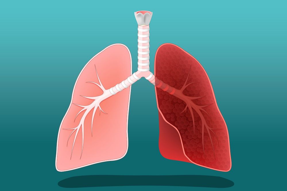
ILD encompasses a broad range of respiratory disorders characterized by inflammation and fibrosis of the interstitium — the delicate network of tissue that surrounds the alveoli in the lungs. This scarring process can significantly impede lung function, leading to a host of respiratory challenges.
The causes of ILD are numerous, and the signs and symptoms are vague, often mimicking other disorders. This makes it extremely difficult to diagnose diseases in this category. Without the correct diagnosis, we cannot treat our patients appropriately.
The name game
ILD is classified based on etiology. When no etiology can be determined, the disease is termed idiopathic.
Types of ILD include:
- Connective tissue disease-associated ILD
- Hypersensitivity pneumonitis
- Drug-induced ILD
- Post-infectious ILD
- Idiopathic interstitial pneumonias
Connective tissue disease-associated ILD is one of the most serious pulmonary complications of connective tissue disease, causing significant morbidity and mortality. Several connective tissue diseases can lead to ILD:
- Rheumatoid arthritis: A systemic inflammatory disease with small joint inflammation
- Systemic sclerosis: A heterogeneous autoimmune disease characterized by immune dysregulation, vasculopathy and progressive fibrosis with multi-organ involvement
- Idiopathic inflammatory myopathies: A group of autoimmune disorders characterized by muscle weakness, rash and autoantibodies
- Dermatomyositis
- Polymyositis
- Antisynthetase syndrome
- Sjogren’s syndrome: Characterized by sicca symptoms, circulating antibodies, lymphocytic infiltration and damage to endocrine glands
- Systemic lupus erythematosus: A chronic autoimmune disease that can affect every organ in the body
- Mixed connective tissue disease: An overlap syndrome associated with muscle weakness, autoantibodies, Raynaud’s phenomenon, dysphagia and arthralgia
Hypersensitivity pneumonitis — also called extrinsic allergic alveolitis — is an inflammatory disease of the lung caused by repetitive inhalation of antigenic agents in a predisposed host. Common agents include:
- Farmer’s lung (most common)
- Grain handler’s lung
- Humidifier or air conditioner lung
- Bird fancier’s lung
- Cheese worker’s lung
- Hot tub lung
Drug-induced ILD is a disease entity that causes significant illness and death. There are reportedly more than 300 drugs that can lead to ILD, including:
- Antineoplastic drugs
- Chemotherapy
- Immunotherapy
- Antibiotics
- Antiarrhythmics
- Immunosuppressants
- Biologic agents
Post-infectious ILD is fibrosis that develops as a result of acute respiratory distress syndrome (ARDS) secondary to pulmonary infections, including COVID-19.
Idiopathic interstitial pneumonias are a subset of ILD with no known etiology. They are further divided into major, rare and unclassifiable idiopathic interstitial pneumonias.
Major idiopathic interstitial pneumonias:
- Idiopathic pulmonary fibrosis (IPF), the most common
- Idiopathic nonspecific interstitial pneumonia (NSIP)
- Respiratory bronchiolitis ILD (RB-ILD)
- Desquamative interstitial pneumonia (DIP)
- Cryptogenic organizing pneumonia (COP), formerly bronchiolitis obliterans organizing pneumonia (BOOP)
- Acute interstitial pneumonia (AIP)
Rare idiopathic interstitial pneumonias:
- Idiopathic lymphoid interstitial pneumonia (LIP)
- Idiopathic pleural-parenchymal fibroelastosis (PPFE)
Unclassifiable idiopathic interstitial pneumonias*
Clinical approach to patients with ILD
When ILD is suspected, the initial approach includes a detailed history from birth to the present and a thorough physical exam, paying special attention to the skin, nails and joints. The preliminary workup involves routine laboratory testing, chest imaging and pulmonary function testing.
Depending on the patient’s exam findings, serology for specific diseases may be ordered. More invasive testing, such as skin, muscle, renal and sinus biopsies, is generally avoided unless necessary.
All cases of ILD should have a team approach. Most institutions have an ILD conference to formally discuss cases before presenting the patient with a diagnosis. Conference members typically include pulmonologists, radiologists, pathologists and rheumatologists. When the diagnosis is still in question, bronchoscopy or surgical lung biopsies can be performed. If there is no definitive radiologic or pathologic diagnosis, the disease is deemed idiopathic.
Medications to treat ILD are designed to slow disease progression; they cannot reverse parenchymal damage. It is important to communicate to patients and their families that, although most ILDs are treatable, treatments come with side effects and limitations. The risks versus benefits should be carefully weighed before starting therapy.
If you suspect that your patient may have ILD, refer them early to a center of excellence or a subspecialist with ILD experience. Most diseases that cause lung parenchymal damage can significantly affect a patient’s physical functioning and shorten life expectancy. Early diagnosis and treatment are crucial to preventing frequent hospitalizations and chronic debility.
In conclusion, the diagnosis of ILD is extremely complicated and labor-intensive.
This article is not intended as a comprehensive overview of ILD but as a starting point for your decision-making process when you suspect a patient has ILD. My goal is to simplify the understanding of this disease entity to improve patient care. Helping providers recognize the complexity of diagnosing ILD may lead to timely referral and immediate treatment.
Learn more about pulmonology services at Northside and find a provider.
*Causes of unclassifiable idiopathic interstitial pneumonia include:
- Inadequate clinical, radiologic or pathologic data
- Major discordance between clinical, radiologic and pathologic findings, which may occur in the following situations:
a. Previous therapy resulting in substantial alteration of radiologic or histologic findings (e.g., biopsy of desquamative interstitial pneumonia after glucocorticoid therapy, which shows only residual nonspecific interstitial pneumonia)111
b. New entity or unusual variant of recognized entity, not adequately characterized by the current American Thoracic Society/European Respiratory Society classification (e.g., variant of organizing pneumonia with supervening fibrosis)222
References:
- Matsushima H, Takayanagi N, Sakamoto T, et al. Pathologic findings both before and after steroid therapy in a case of desquamative interstitial pneumonia. Nihon Kokyuki Gakkai Zasshi. 2001;39:609.
- Lee JW, Lee KS, Lee HY, et al. Cryptogenic organizing pneumonia: Serial high-resolution CT findings in 22 patients. AJR Am J Roentgenol. 2010;195:916.